
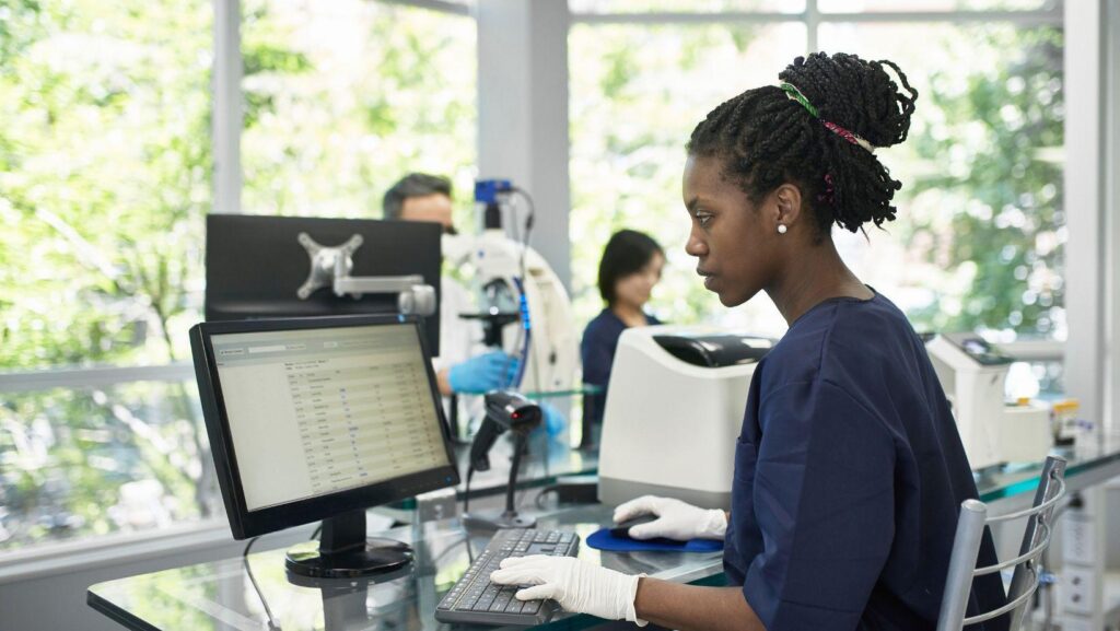
A decade ago, digital pathology was mostly a novelty, something a handful of academic centers experimented with while everyone else stayed comfortably attached to their microscopes. That’s changed. Whole slide imaging has moved from an interesting side project to a real clinical tool, and the labs getting the most out of it are the ones that figured out how to tie it tightly into their laboratory information system rather than running it as a separate, disconnected process.
This piece looks at what that integration actually looks like in practice, and why the connection between digital slides and the LIS matters more than people sometimes expect going in.
What Digital Pathology Actually Involves
At a basic level, digital pathology means scanning glass slides into high-resolution digital images that pathologists can review on a screen instead of, or in addition to, a physical microscope. A slide scanner captures the image, and that file needs to end up somewhere a pathologist can actually access it alongside the rest of the case information.
That last part is where things get complicated. A scanned slide floating around in a separate image management system, disconnected from the case record, patient history, and grossing notes, doesn’t save anyone time. It just moves the friction somewhere else.
Why the LIS Connection Matters So Much
The value of digital pathology shows up fully only when the images are tied directly into the case within the laboratory information system. When that connection is done right, a pathologist opens a case and sees the grossing description, prior history, and the digital slide all in one place, without needing to jump between separate platforms or manually match up file names to case numbers.
Labs that skip this integration often end up with pathologists doing exactly that kind of manual matching, hunting down the right image file, cross-referencing it against a case number, and hoping nothing gets mixed up along the way. In a specialty where a mismatched slide can lead to a wrong diagnosis, that’s not a small risk.
Supporting Remote and Distributed Sign-Out
One of the biggest practical shifts digital pathology has enabled is remote case review. Pathologists no longer need to be physically present in the same building as the physical slides to sign out a case. A subspecialist working from a different office, or even a different state, can review a digital slide and complete sign-out as long as the system supports it properly.
This has real implications for how pathology groups staff their operations. A multi-site group can route a complex dermatopathology case to the one subspecialist in the network best equipped to handle it, rather than being limited to whoever happens to physically be in that building that day. None of this works smoothly without an LIS built to handle remote access, proper case routing, and secure image delivery as core functionality rather than an afterthought bolted onto an older system.
AI-Assisted Review and Where the LIS Fits In
Digital slides have also opened the door to AI-assisted analysis, tools that can flag areas of interest on a slide, assist with quantification tasks like counting mitotic figures, or support certain screening workflows. These tools are becoming more common, particularly in high-volume areas like prostate and breast pathology.
The LIS plays a coordinating role here too. Rather than pathologists needing to run a separate AI tool outside their normal workflow, well-integrated systems surface AI-generated annotations or flags directly within the case review interface. This keeps the pathologist’s attention in one place instead of splitting focus across multiple disconnected tools, and it keeps a record of what the AI flagged as part of the permanent case documentation, which matters for both quality assurance and, increasingly, regulatory expectations around AI use in diagnostics.
Storage, Bandwidth, and the Less Glamorous Technical Realities
It’s worth being honest about the less exciting side of digital pathology too. Whole slide images are large files, often multiple gigabytes per slide depending on scan resolution, and a busy AP lab can generate a lot of them daily. That creates real storage and bandwidth demands that a laboratory information system, or the infrastructure supporting it, needs to be built to handle.
Cloud-based platforms tend to manage this more gracefully than older on-premises systems, since scaling storage and bandwidth in the cloud is a matter of provisioning rather than physically expanding server infrastructure. Labs evaluating a digital pathology rollout should ask pointed questions about how their LIS handles image storage at scale, not just how it performs with a handful of demo slides during a sales pitch.
Quality Control in a Digital Workflow
Digital pathology introduces some quality considerations that didn’t exist in a purely glass-slide world. Scan quality can vary, and a poorly scanned slide can obscure diagnostically important detail just as easily as a poorly cut physical section can. A well-built LIS supports quality control checkpoints specifically for scanned images, flagging scans that fall outside expected quality parameters before they ever reach a pathologist for review.
This kind of built-in checkpoint matters because catching a bad scan early is a lot less costly than catching it after a pathologist has already spent time reviewing a case, only to discover the image quality wasn’t sufficient for a confident diagnosis.
The Bigger Picture
Digital pathology isn’t really about replacing the microscope, at least not yet for most labs. It’s about adding flexibility, enabling remote consultation, supporting AI-assisted tools, and creating a more complete digital record of every case. None of that works particularly well if the imaging side and the case management side are running as two separate systems that happen to be used by the same lab.
The labs seeing the most benefit from digital pathology tend to be the ones that treated the LIS integration as a core part of the rollout from the beginning, not something to figure out after the scanners were already installed. Getting that connection right is really what determines whether digital pathology becomes a genuine workflow improvement or just an expensive parallel system nobody fully trusts.